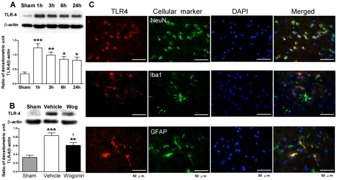
Figure 7. Effects of 40 mg·kg−1 wogonin on TLR4 expresssion.
(A) Representative immunoblots of toll-like receptor (TLR)-4 protein in the ipsilateral hemisphere from untreated injured mice. Bar graphs of densitometry analysis of the protein bands showed increased TLR4 levels at 1, 3, 6 and 24 h post-injury (n = 5 mice/time point). (B) Representative immunoblots showing TLR-4 protein in the ipsilateral hemisphere from a sham-injured control, a wogonin-treated injured mouse, and a vehicle-treated injured mouse at day 1 post-TBI. Bar graphs of densitometry analysis of protein bands showed a significant decrease in TLR4 protein levels in the ipsilateral hemispheres of wogonin-treated mice at day 1 post-TBI compared with vehicle-treated mice. (C) Identification of TLR4- positive cells 1 day post-injury in the peri-contusion margin by immunofluorescence labeling. TLR-4 immunoreactivity is shown in red, and immunolabeling of NeuN (a cell marker for neurons), anti-ionized calcium binding adaptor molecule 1 (Iba1, a cell marker for microglia), or GFAP (a cell marker for astrocytes) is shown in green. Yellow labeling indicates co-localization. TLR4 was co-localized in neurons and astrocytes, and poor colocolization was observed in microglia. Sections were stained with DAPI (blue) to show all nuclei. The scale bar is 50 µm. Values are reported as means ± SEM; *P<0.05, **P<0.01, ***P<0.001 versus sham controls, and † P<0.05 for wogonin-treated versus vehicle-treated TBI mice (n = 7 mice/group).

