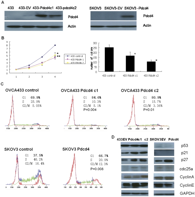
Figure 1. PDCD4 inhibited cell proliferation and cell cycle progression through up regulation of p21 and p27.
(A) PDCD4 over-expressing stable clones 433-PDCD4c1, 433-pdcd4c2, and SKOV3-PDCD4, were established in ovarian cancer cells OVCA433 and SKOV3. 433-EV and SKOV3-EV: GFP over-expressing empty vector stable clones for control. (B) 433 PDCD4c1 and 433 PDCD4c2 exhibited significant slower proliferation rate compared with control (*p<0.01 and **p<0.05, respectively) according to XTT (left panel) and clonogenic assay (p<0.05) (right panel). (C) PDCD4 induced cell cycle arrest at G1 stage. Representative data are from one of the three independent experiments. The percentages of cells that were in G1 stage for OVCA433 c1 and c2 were 84.6% (±2.1%) and 80.9% (±2.2%), respectively. Both were significantly higher compared with control (69.9%±3.1%) (p = 0.004 and P = 0.01, respectively). The percentage of cells that was in G1 stage for SKOV3-PDCD4 was 66.7% (±1.8%), which was significantly higher compared with control (37.5%±2%) (p = 0.008). (D) PDCD4 over-expressing stable clones as well as control empty vector stable clones were maintained in MEM with 10% FBS, and then harvested for protein extraction. Protein expressions of a panel of cell cycle regulators including p53, p21, p27, cdc25a, cyclinA and cyclinE were analyzed. The intensity of the band was determined by densitometric scanning. The quantification of the bands was presented in Data S1. GAPDH was included as internal loading control. Three independent experiments were performed.

