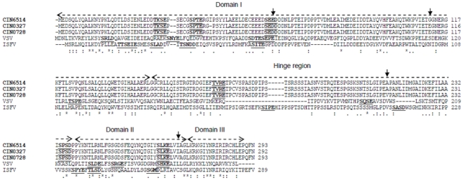
Figure 4. Alignment of P protein of representative CHPV isolates (indicated in bold) with closely related vesiculoviruses VSV and ISFV.
All putative phosphorylation sites are underlined. The different domains defined for the P protein of VSV are indicated by two-headed dashed overhead arrows. The mutations within the CHPV isolates are indicated by downward arrows.

