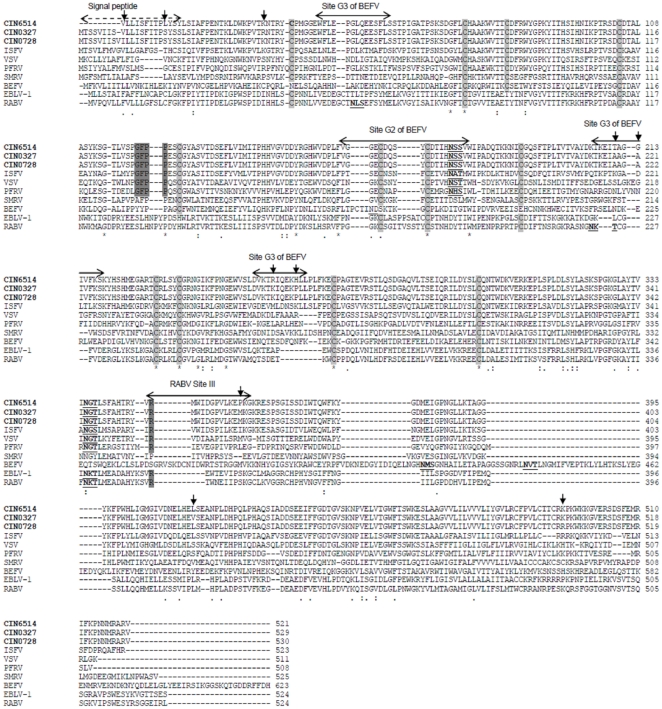
Figure 6. Alignment of G protein of representative CHPV whole genome isolates (in bold) with other representative rhabdoviruses.
BEFV antigenic sites and RABV antigenic sites are indicated by arrows above the sequence. Functionally important motifs are highlighted in grey and putative glycosylation sites are underlined. The signal peptide is indicated by dashed arrow. Highly conserved cysteine residues are highlighted in faint grey. Mutations within CHPV isolates are indicated by downward arrows.

