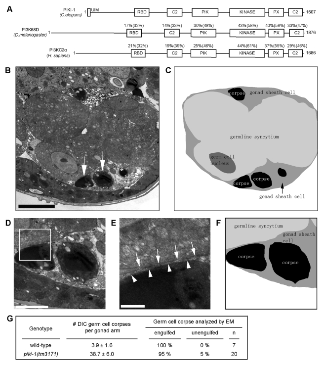
Figure 2. In the gonad of piki-1(tm3171) mutants, apoptotic germ cells are engulfed but fail to be degraded.
(A) Domain structure of PIKI-1 and its orthologs in other species. Percentage indicates amino acid identity (similarity in parentheses) of each domain between PIKI-1 and its orthologs; UIM, ubiquitin-interacting motif; RBD, Ras-binding domain; C2, protein kinase C conserved region 2; PIK, phosphoinositide 3-kinase, accessory domain; Kinase, phosphoinositide 3-kinase, catalytic domain; PX, PhoX homologous domain. (B–C) A cross-section transmission electron microscopy (TEM) image of a distal gonad arm in a piki-1(tm3171) adult hermaphrodite (B) and its corresponding traces of membranes (C). Cell identities are labeled. White arrows in (B) indicate two engulfed cell corpses. The black arrow in (C) indicates a gonadal sheath cell. Scale bar: 5 µm. (D–F) Enlarged images of germ cell corpses indicated by arrows in (B) and their surrounding environment inside a sheath cell are displayed in (E). (F) Schematic diagram of (D). (E) Further enlarged image of the framed region in (D). White arrows and arrowheads mark the plasma membranes of gonadal sheath cells and germ cell corpses, respectively. Scale bars: 2 µm in (D); 0.5 µm in (E). (G) Percentage of engulfed and unengulfed germ cell corpses quantified by TEM. Data of wild-type animal are from [20]. n, number of germ cell corpses analyzed.

