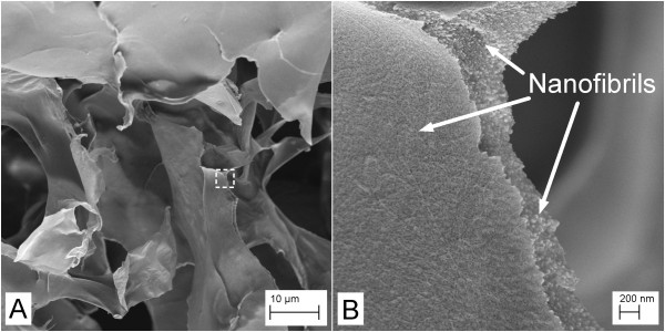
Figure 3.
FESEM images of a gel prepared from nanofibrils mixed with pNIPA particles. Images acquired at relatively (A) low and (B) high magnifications. The image in (B) has been acquired from the area marked with a dotted rectangle in the image in (A). Note the nano-sized fibrils forming the network structure.

