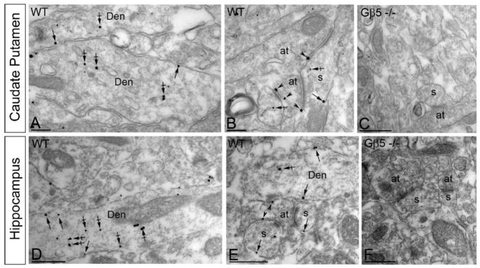
Figure 2. Subcellular localization of Gβ5 in the striatum and hippocampus.
Electron micrographs show immunolabeling for Gβ5 in different neuronal compartments of WT mice, as detected using a pre-embedding immunogold method. In striatum (A-C), and hippocampus (D-F) immunoparticles for Gβ5 were detected postsynaptically along the somatic plasma membrane (arrows), and intracellular sites (crossed arrows) of dendritic shafts (Den) and dendritic spines (s. In addition, immunoparticles for Gβ5 were detected at presynaptic sites, along the extrasynaptic plasma membrane (arrowheads) of axon terminals (at) establishing excitatory synapses with dendritic shafts or dendritic spines (s). Immunoreactivity for Gβ5 was completely absent in Gβ5−/−samples (C,F). Scale bar: 0.2 μm.

