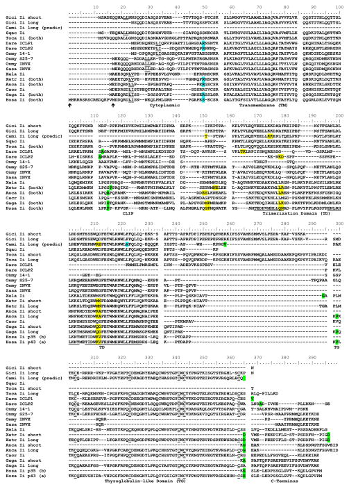
Figure 2. Amino acid alignment identifies nurse shark and other chondrichthian Ii when compared to other vertebrate sequences.
Yellow highlighting (spanning two residues) indicates phase zero, green highlighting indicates phase one and blue highlighting indicates phase two intron positioning. Genbank accession numbers are shown in Supplemental Table 2. Endosomal targeting motifs (L-L/I/V) are underlined, as are the six conserved cysteines of Tg-like domains and (in the human sequences) the three alpha helices of the trimerization domain. Asparagine-linked glycosylation motifs (N-X-S/T, X≠P) are in italics. Also in italics are polar residues corresponding to the hydrophilic patch of the transmembrane domain implicated in Ii trimerization.

