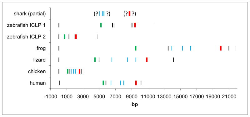
Figure 4. Exon-intron organization and relative size similar from shark to man.
Exons encoding the transmembrane domain are shown in green, the exons approximately encoding the three alpha-helices of the trimerization domain are shown in blue, and the Tg domain in red. Question marks denote missing data from elephant shark scaffolds, exon content in each set of parentheses is on one unmapped scaffold. Drawn to scale, distance in base pairs is shown at bottom.

