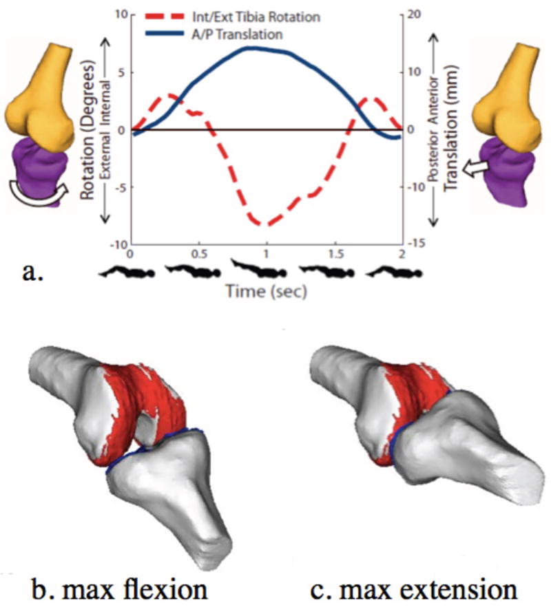
Figure 3.

3D joint kinematic data (a) and models (a,b,c) derived from Cine-PC MR images (TR/TE: 6.8/3.3 msec) of a healthy knee acquired in 5:36 minutes under a load similar to that of walking. External tibial rotation and anterior tibial translation can be visualized from extension to 37° of flexion (a) and when coupled with segmented bone and cartilage models, can be used to demonstrate contact and motion of the tibio-femoral joint throughout a cycle of flexion (b) and extension (c). (Reproduced from Bradford R, Johnson K, Wieben O and Thelen D. Dynamic imaging of 3d knee kinematics using PC-VIPR. In: Proceedings of the 19th Anuual Meeting of ISMRM, Montréal, Québec, Canada, 2011 (Abstract 3178), with permission.)
