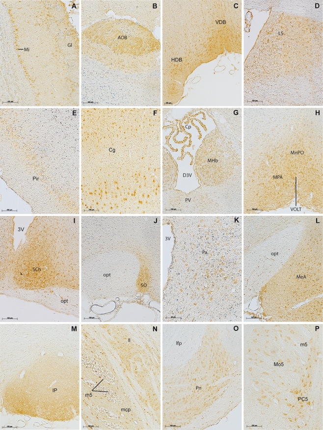
Fig. 3.
a, b. Olfactory system. Alarin-LI-positive cells were detected in the mitral cell layer, the glomerular cell layer, and the accessory olfactory bulb. c, d. Basal forebrain. The nuclei of the vertical and horizontal limb of the diagonal band and the lateral septal nucleus revealed alarin-LI. e, f. In the cortex alarin-LI-positive cells were detected in the piriform and cingulate cortex. g In the thalamic area and third ventricle, the choroid plexus and medial habenular nucleus show strong alarin-LI and weak alarin-LI-positive cells in the paraventricular thalamic nucleus. h–j Prepotic area. Intense alarin-LI-positive cells and diffuse labeling are detected in the medial preoptic area, the median preoptic nucleus, the vascular organ of the lamina terminalis, the suprachiasmatic nucleus, the supraoptic nucleus, and in the lateral hypothalamic area. Only single cells are detected in the optic tract. k Alarin-LI in magnocellular secretory cells of the paraventricular hypothalamic nucleus. l Intense alarin-LI of cells detected in the medial amygdala and small single cells in the optic tract. m Diffuse alarin-LI in the interpeduncular nucleus. n Small single cells positive for alarin-LI indicated in the motor root of the trigeminal nerve and middle cerebellar peduncle. In the lateral lemniscus, cell bodies revealed to be positive for alarin-LI. o Strong alarin-LI of medium-sized cells detected in pontine nuclei. p Alarin-LI-positive neurons in the motor trigeminal nucleus, the motor root of the trigeminal nerve, and the parvicellular motor trigeminal nucleus. q In the pond, the locus coeruleus showed alarin-LI-positive neurons. r Alarin-LI-positive neurons in the nucleus of the trapezoid body. s In the chochlear nucleus, small single cells were alarin-LI positive. t Alarin-LI-positive neurons in the facial nucleus. Abbreviations of nuclei are given in Table 1. Abbreviations not included in Table 1: 3V third ventricle, D3V third ventricle dorsal part, lfp longitudinal fasciculus of the pons, scale bars = 100 μm


