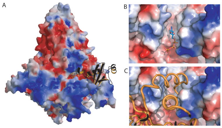
Fig. 3. Intermolecular contact in the p110α /p85 heterodimer crystal.
(A) Molecular surface of the PI3K colored as electrostatic charges showing the Ras binding domain of a neighboring molecule in the crystal (orange ribbon with black back-side) bound in the kinase domain active site. (B) Molecular surface colored as electrostatic potential showing the ATP (ball-and-stick representation) bound to PI3Kα . (C) Molecular surface colored according to the electrostatic potential showing the helix-loop-helix motif of the RBD of the neighboring molecule in the ATP binding site.

