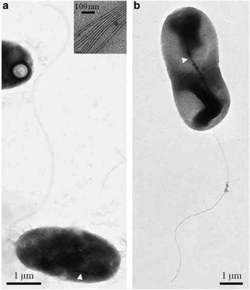
Figure 2.
TEM images of negatively stained (1% uranyl acetate) cells of the magnetotactic Gammaproteobacteria. (a) TEM image of cells of strain BW-2 showing chain of magnetite magnetosomes (at white arrow) and a polar bundle of flagella. Inset shows that the flagellar bundles consist of seven individual flagellum. (b) TEM of cells of strain SS-5 showing chain of magnetosomes (at white arrow) and a single polar flagellum.

