
Figure 2.
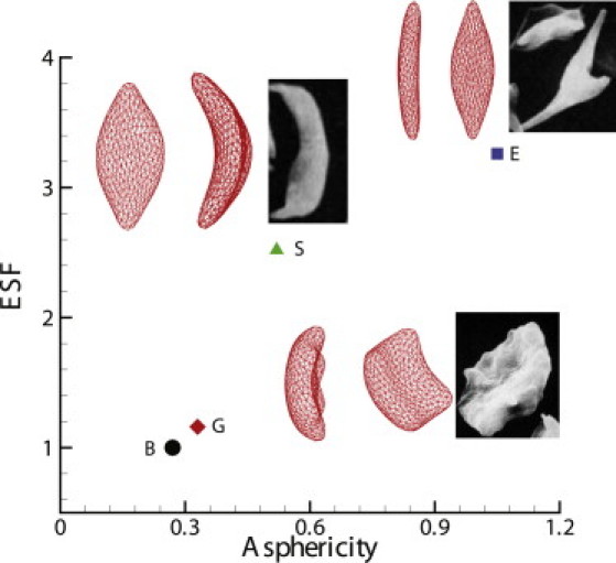
ASF and ESF for the different shapes of sickle cells. Labels G, S, and E represent the granular, sickle, and elongated shapes of the sickle cells, respectively, and the inset shows their morphologic projections on the x-z and x-y planes. The inset images represent experimental observations for different morphologic states of deoxygenated SS-RBCs by SEM (reproduced with permission from Kaul and Xue (10)). The label B corresponds to the original biconcave shape, the morphological projection of which is shown in Fig. 1.