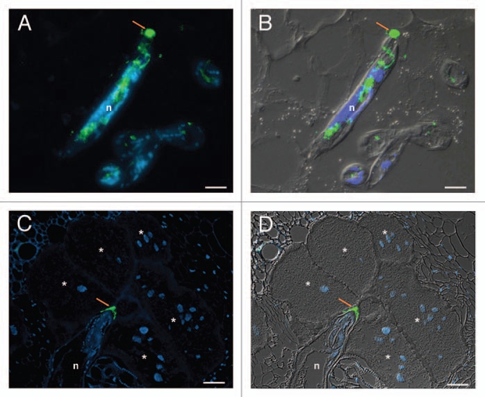
Figure 1.

Immunodetection of Meloidogyne incognita secreted proteins during parasitism. (A and B) Localization of secreted CBM2-bearing proteins in the apoplasm (arrow) during nematode migration in Arabidopis thaliana root sections. (C and D) Localization of secreted MAP-1 in the apoplasm accumulating along the giant cell wall (arrow) by sedentary nematode in tomato (Solanum lycopersicum) root sections. Micrographs (A and C) are overlays of an Alexa-488 fluorescence (green) and DAPI-stained nuclei (blue). Micrographs (B and D) are overlays of an Alexa-488 fluorescence (green), DAPI-stained nuclei (blue) and differential interference contrast (grey). n, nematode. *, giant cells. Scale bars = 10 µm. Micrographs (A and B) are from Vieira et al. 2011.23
