
Figure 3.
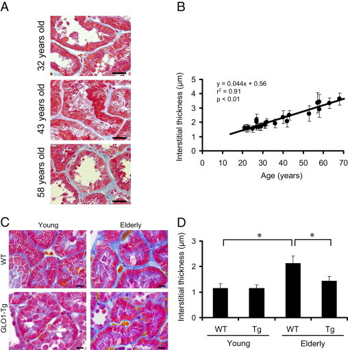
Interstitial thickening was demonstrated to be an age-related morphological change in human and rat kidney. A: Representative figures of human renal biopsy. Specimens were Azan-stained. Proximal tubular epithelial cells stain red and interstitium stains blue. The interstitial areas were thickened in an age-dependent manner. Original magnification = ×600. Scale bars: 20 μm. B: Correlation between age and interstitial thickness. The thickness of renal interstitia (blue) was measured under high-power magnification (×600). Each dot on the univariate linear regression line represents mean thickness (μm); error bars indicate ± SD. The calculation method is described under Materials and Methods. C: Light micrographs of rat renal cortex. Formalin-fixed specimens (3 μm thick) were stained with Masson's trichrome staining. Proximal tubular epithelial cells with brush borders stain red; interstitial areas stain blue (as for the human renal biopsy). The interstitial areas were thickened with age. This age-dependent thickening was attenuated by GLO1 overexpression. Original magnification = ×600. Scale bars: 10 μm. D: Quantitative histological analysis indicates that GLO1 overexpression ameliorated age-dependent interstitial thickening. *P < 0.01; **P < 0.05.