
Figure 1.
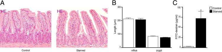
Morphological and functional changes in the starved intestine. A: H&E staining of ileum from control (left panel) and starved (right panel) mice showed no apparent changes in intestinal architecture. B: Villus length and crypt depth were similar in the ileum of starved mice compared with controls. C: Assessment of ileal permeability demonstrated a significant increase in plasma 4.4-kDa FITC-dextran of starved mice compared with controls. *P < 0.05. The histological features are representative for all tissue samples studied (n = 15 per group).