
Figure 3.
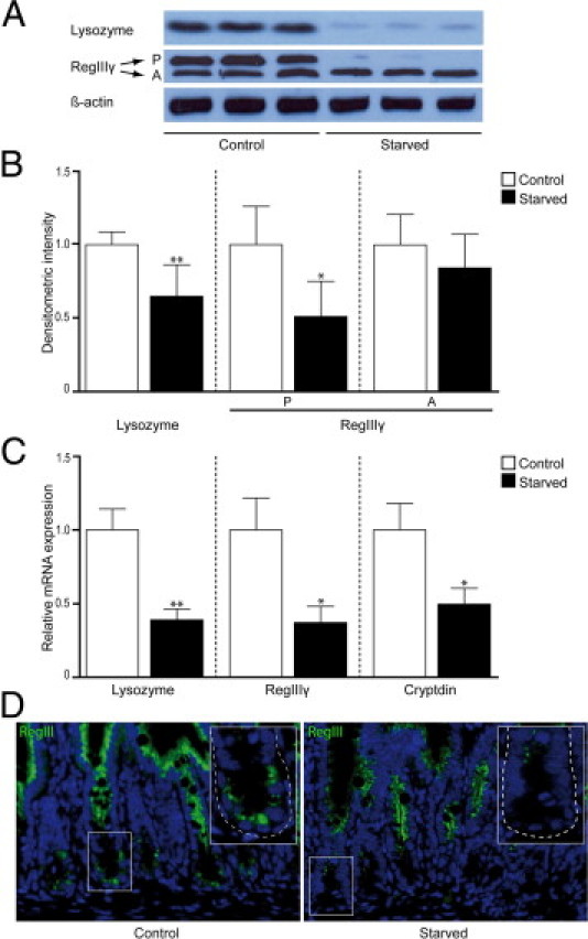
Protein and mRNA levels of Paneth cell antimicrobial proteins are decreased after 48 hours of starvation. A: Western blot analysis showed decreased band density for lysozyme (15 kDa) and the precursor (P) form of RegIIIγ (16.5 kDa) in the ileum of starved mice compared with control mice, whereas no change was observed in the active form (A) of RegIIIγ (representative bands of three mice per group are shown). β-Actin was used to correct for equal protein loading (n = 12 per group). B: Quantification of Western blots demonstrated a significant decrease in both lysozyme (*P < 0.01) expression and expression of the precursor form of RegIIIγ (**P < 0.05). C: qPCR analysis showed a 2.5-fold decrease in lysozyme gene expression (*P < 0.01), a 2.7-fold decrease in gene expression of RegIIIγ (**P < 0.05), and a twofold decrease in cryptdin (**P < 0.05) gene expression after starvation (n = 15 per group). D: Immunofluorescence for RegIIIγ in ileum from control (left panel) versus starved (right panel) mice showed an overall reduced expression of RegIIIγ in fasted animals, as well as in Paneth cells specifically. Insets: Magnifications of single crypts from both control and starved mice. The histological features are representative for all tissue samples studied (n = 15 per group).