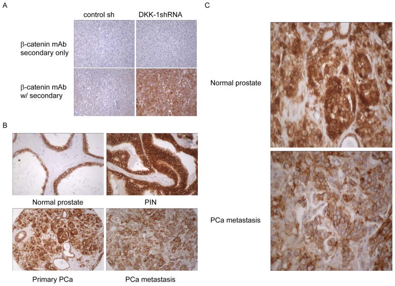
Figure 3. Totalβ-catenin and DKK-1 concomitantly decrease during PCa progression.
A. Specificity of β-catenin antibody. Serial sections from paraffin embedded tumor tissue produced byβ-catenin− PC-3 shRNA control transfected PCa cells or β-catenin+ PC-3 DKK-1shRNA transfected PCa cells were incubated using the protocol described in the Materials and Methods section with secondary only or primary antibody to β-catenin. B. A progression TMA was stained for β-catenin using routine immunohistochemistry. Shown are tissue cores that represent the median of β-catenin percent expression of normal prostate, PIN, primary PCa and PCa metastases presented in Figure 4A (200x). Brown color indicates β-catenin staining. C. High power (400x) magnification of primary PCa and metastatic lesions showing reduced nuclear β-catenin staining.

