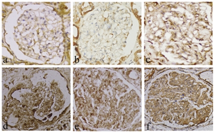
Figure 4. OTUB1 expression in glomerular during some golmerulonephritides was detected by immunohistochemistry.
(a) Normal glomerulus, there is few positive staining in golmerulus; (b) minimal change disease; (c) membranous glomerulonephritis, there is minor positive in the mesangium at (b) and (c); (d) IgA nephropathy, moderate positive in the mesangium; (e) acute proliferative glomerulonephritis; (f) lupus nephritis. The increased positive staining was seen in mesangium at (d), (e) and (f). In addition, the tubular epithelial cells are partially diaminobenzidine positive. Hematoxylin is used as the nuclear counterstain. (ABC immunochemistry, ×100).

