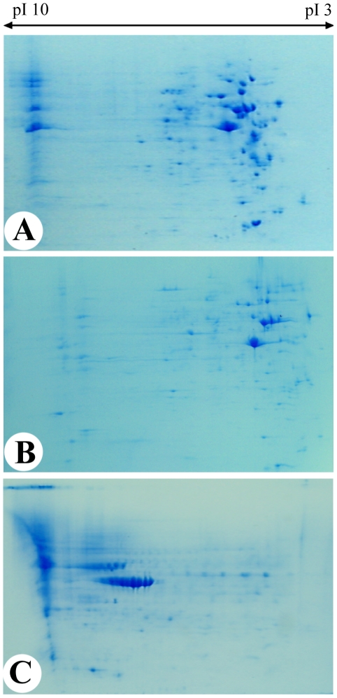
Figure 1. Two dimensional electrophoresis gels.
The gels are over the broad pI range of 3 to 10 for AP cells (1A), PP cells (1B) and FP cells (1C). Note that most of the proteins in AP cells and PP cells were in the low pI region of the gel. AP cells, antlerogenic periosteum cells; PP cells, pedicle periosteum cells; and FP cells, facial periosteum cells.

