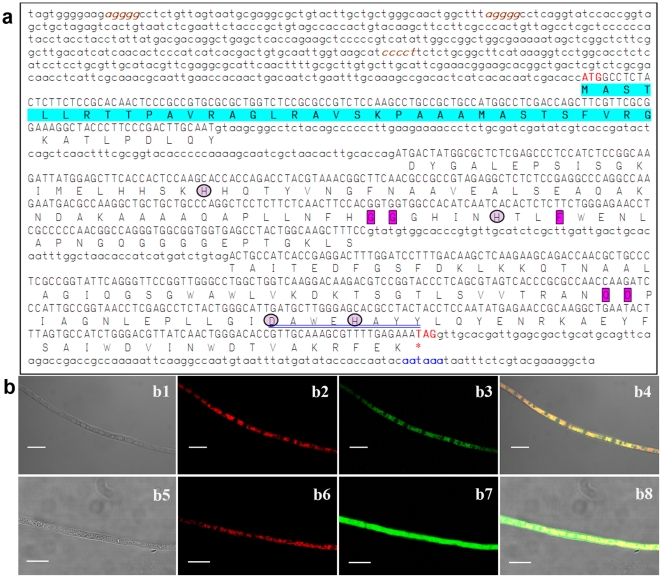
Figure 1. The features of BbSod3 encoding mitochondrial MnSOD in B. bassiana.
(a) The nucleotide and deduced protein sequences of BbSod3. The uppercase DNA fragment is a 693-bp ORF encoding a protein of 230 amino acids while the lowercase fragments are ORF-flanking regions. Located in 5′ UTR and 3′ UTR are three putative stress-response elements (brown and italisized) and a putative polyadenylation signal (blue), respectively. Note that the first 34 amino acids (highlighted) of the deduced protein were predicted as a mitochondria-targeted signal peptide. The framed residues are the Parker and Blake signatures typical for the Mn-SOD family and the circled residues are metal-binding sites. The sequence double-underlined in blue represents the consensus pattern DXWEHXXY for the Fe/Mn-SOD superfamily. (b) Intracellular localization of BbSod3. Mycelia of transgenic strains expressing the fusion BbSod3signal::eGFP (b1–b4) and the signal-free eGFP (b5–b8, control) were stained with the mitochondria-probing stain MitoTracker® Red, emitting the red (stain) and green (eGFP) fluorescences. The differential interference contrast image (b1) and the same image labeled with the stain (b2) and the expressed fusion (b3) overlapped very well, forming the merged image (b4) whose color pattern (entirely brown-yellow in reticulum components) indicates the mitochondrial target of the fused signal peptide, whereas the merged control image (b8) showed more green than yellow. Scale bars: 10 µm.

