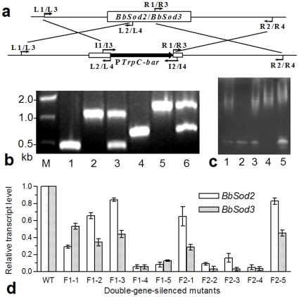
Figure 3. Knockout and complement of BbSod2 and BbSod3 and RNAi double silence of both enzymes.
(a) Diagram for the knockout constructs of BbSod2 and BbSod3 (see Table 1 for the used primers). (b) Detection of the disrupted and complemented BbSod2 and BbSod3 fragments by PCR with paired primers I1/I2 (Lanes 1–3: WT, ΔBbSod2 and ΔBbSod2/BbSod2) and I3/I4 (Lanes 4–6: WT, ΔBbSod3 and ΔBbSod3/BbSod3). (c) SOD active bands on the NBT-stained gels of WT (Lane 1) and mutants (Lanes 2–5: ΔBbSod2, ΔBbSod2/BbSod2, ΔBbSod3 and ΔBbSod3/BbSod3, respectively). (d) RNAi double silence. Relative transcript levels of BbSod2 and BbSod3 in 10 RNAi mutants of the fused genes F1 and F2 were assessed via qRT-PCR. Both fusions were constructed with BbSod2 ORF and partial BbSod3 ORF. Note that the desired double-gene silence was achieved in the mutants F1-4, F2-2 and F2-4. Error bars: SD of the mean from three replicates.

