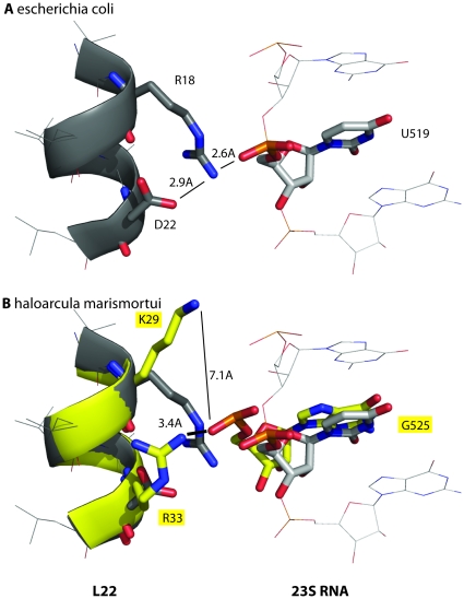
Figure 4. Structural evidence explains the residue distributions for triplet U519/R18/D22.
In E.coli, the values of RNA U519/L22 R18/L22 D22 are U, Arg (R) and Asp (D), respectively. A hydrogen bond network in E.coli goes from the side chain of D22 to the side chain of R18 to the phosphate atom of U519. (Figure 4A), and explains the tight coupling seen in the distribution (Figure 3). The triplet from the archeon haloarcula marismortui represents a shift from pyrimidine to purine, with the values of U519/R18/D22 at G, Lys (K) and Arg (R), respectively. A structural alignment of the crystal structure from both species reveals that the hydrogen bonds are broken when the RNA is a purine and the residues farther apart (Figure 4B). This data suggests that the change in packing to accommodate a larger RNA side chain influences the packing between the L22 and 23S protein in such a way that this hydrogen bond network is broken.

