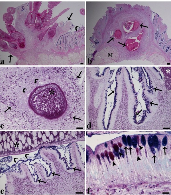
Figure 2.

Transverse section through the intestine of a tench infected with several Monobothrium wageneri. (a) Transverse section through the intestine of a tench infected with several Monobothrium wageneri showing a pronounced inflammatory response surrounding the scoleces and a marked lack of epithelia at the site of tapeworm attachment (arrows). Note the presence of intact epithelia (curved arrow) in close proximity to the nodule, scale bar = 200 μm. (b) Focal attachment of several M. wageneri penetrating the intestine of a tench as far as the muscularis. An intense host cellular reaction around the scoleces is visible (arrows), M = muscularis, scale bar = 200 μm. (c) Scolex of M. wageneri (asterisk) is surrounded by numerous granulocytes (arrow heads) and collagenous fibres (curved arrows), scale bar = 50 μm. (d) A high number of mucous cells (arrows) are visible within the epithelia in the immediate vicinity of the scolex (asterisk). Note the intense recruitment of granulocytes (arrow heads) within the sub-mucosa, scale bar = 100 μm. (e) The villi adjacent to the body of the cestode (asterisk) are seen to be covered with a blanket of mucus (curved arrows) and possess a high number of mucous cells (arrows). Numerous granulocytes (arrow heads) are evident within the sub-mucosa, scale bar = 100 μm. (f) Alcian blue/PAS stained mucous cells close to the site of cestode attachment. The arrows indicate that most mucous cells stain positively for acid glycoconjugates whilst the arrow heads indicate that a lower number of mucous cells stain for the presence of mixed glycoconjugates, scale bar = 10 μm.
