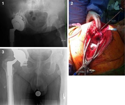
Fig. 1.
1 Pre-operative radiograph. The patient’s acetabular component has shifted, and the THA has recurrent anterior dislocations. 2 Intra-operative photo showing custom cage-cup implanted. Probe is pointing to intact external rotator complex. 3 Post-operative radiograph. The patient’s cage-cup has been implanted according to pre-operative plan. Additional offset was used to adjust for patient’s prior instability

