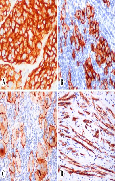
Fig. 1.
KBA62 in melanocytic neoplasms. A. Clusters of nevus cells show strong cytoplasmic positivity. B. Nodal melanoma metastasis shows membranous, cytoplasmic and Golgi-zone staining pattern. C. Pleomorphic metastatic melanoma shows prominent membrane and variable cytoplasmic and golgi-zone staining. D. Bundles of desmoplastic melanoma cells are KBA62-positive.

