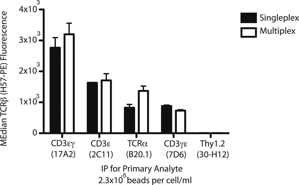
Figure 5.
Inter-bead independence is evident by comparing single- to multiplex IP-FCM. IP beads specific for the primary analyte indicated on the x-axis were incubated with TOT-1 cell lysate. For this experiment, 2 × 10³ beads were incubated with cell lysate consisting of 1.75 × 10⁶ cells/0.02 mL lysis volume (2.3 × 10⁻⁵ beads per cells/mL). Captured TCR complexes were stained for co-associated TCRβ, with resulting fluorescence depicted on the y-axis. Black bars, IP-FCM performed in singleplex using the single IP bead indicated. White bars, IP-FCM performed in multiplex using the combination of the five IP beads listed.

