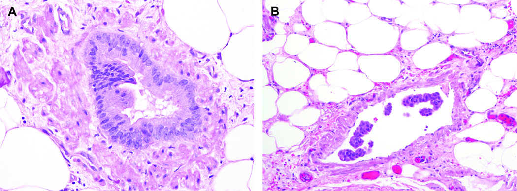
Figure 1.
Representative images of PanIN-like vascular invasion and conventional vascular invasion. When the invasive neoplastic cells form complete luminal structures inside of the blood vessels, we classified the case as having “PanIN-like vascular invasion” (A). When the neoplastic cells were inside of the blood vessel but did not form complete lumina, we classified the case as having “conventional vascular invasion” (B).

