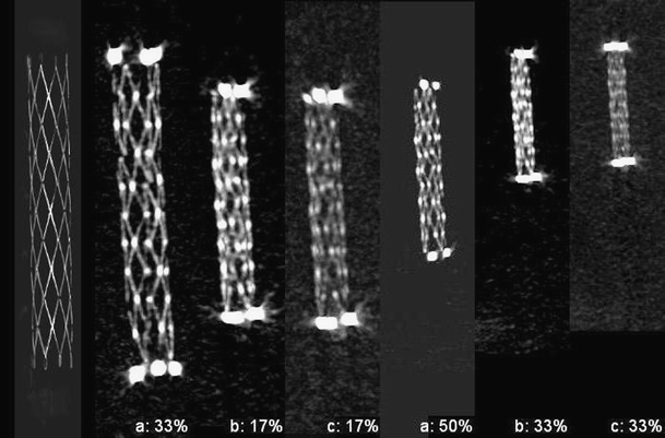
Fig. 1.

A Wingspan nitinol stent. A comparison between a the high-resolution/high-contrast case, b standard resolution/high-contrast case, and c the standard resolution/standard contrast case. In all cases, an intra-tube contrast agent of 31 HU was used. The relevant reconstruction (sub) volumes are denoted as 50% and 33% for the HIRES case. For the standard resolution, 17% and 33% images are shown. For comparison, a contrast inverted optical image is shown on the far left
