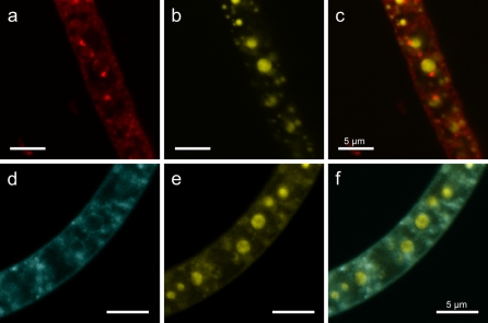
Fig. 2.
Fluorescence of Beggiatoa strain 35Flor filaments obtained by dual staining with DAPI for polyphosphate and Nile Red or MDY-64 for lipid detection. a Lipid layers of spherical structure and different sizes are visible by staining with Nile Red. b In the same filament, stained with DAPI, polyphosphate inclusions of different sizes are visible by a yellow fluorescence signal. c An overlay of the Nile Red and DAPI fluorescence shows the existence of lipid layers for most of the large and some of the small polyphosphate inclusions. d Staining with MDY-64 reveals the same pattern of lipid layers as for Nile Red staining. e Polyphosphate inclusions of different sizes. f The overlay of MDY-64 and DAPI fluorescence reveals that most polyphosphate inclusions are enclosed by a lipid layer indicating a membrane. Note The detected internal lipid layers do not exclusively surround polyphosphate inclusions. Scale bars represent 5 μm

