Fig. 2.

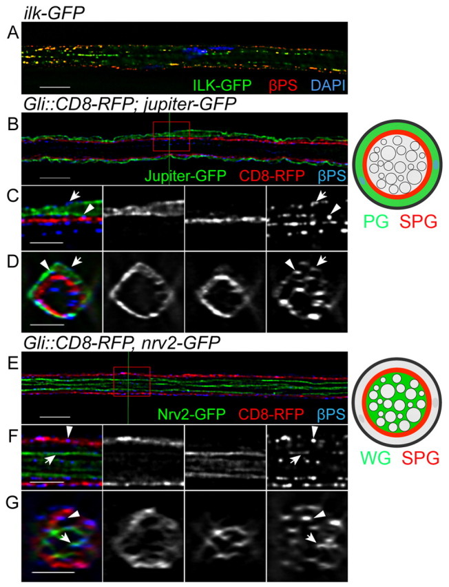
βPS integrin is expressed in the peripheral glia layers. Immunolabeling for the βPS integrin subunit in Drosophila third instar nerves expressing either Ilk-GFP or different glial markers in single 0.2 μm z-sections. (A) βPS (red), Ilk-GFP (green) and DAPI (blue) labeling. Both βPS and Ilk form puncta and are associated with each other in the peripheral nerve. (B-G) βPS labeling in the different glial subtypes: PG using Jupiter-GFP (B-D, green), SPG using Gli-GAL4::CD8-RFP (B-G, red) and WG using Nrv2-GFP (E-G, green). The labeled glial layers are also illustrated to the right. The red boxes in B and E were digitally expanded as shown in C and F, respectively. The green lines in B and E indicate the positions of the orthogonal sections in D and G, respectively. βPS integrin was found in the PG outer membrane (C and D, arrow), the PG-SPG boundary (C and D, arrowhead), the SPG inner membrane (F and G, arrowhead) and the WG membrane (F and G, arrows). Scale bars: 10 μm in A,B,E; 5 μm in C,D,F,G.
