
Fig. 7.
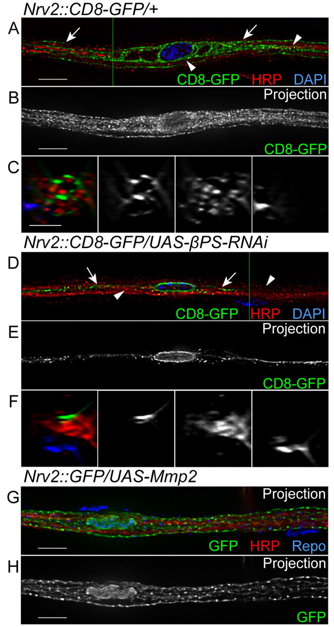
Expression of βPS-RNAi in the wrapping glia. Nrv2-GAL4 was used to express CD8-GFP (green) and βPS-RNAi in the WG. Anti-HRP and DAPI labeling were used for axons (red) and nuclei (blue), respectively. The labeling and phenotypes observed in the WG are illustrated to the right. (A,D) Single 0.2 μm z-sections; (B,E,G,H) projections of the entire z-stack; (C,F) orthogonal sections at the positions indicated by the green lines. (A-C) In a control nerve, CD8-GFP surrounded the DAPI-labeled glia nucleus (blue) and extended along the nerve to fill complex processes (arrows) in between the HRP labeling (red, arrowheads). (D-F) In an Nrv2::βPS-RNAi nerve, CD8-GFP was found only in a single process that extended along the labeled axons (red, arrowheads). Arrows point to small projections and puncta of CD8-GFP. (G,H) In an Nrv2::Mmp2 nerve, the CD8-GFP-labeled processes of the WG appeared normal and spread throughout the labeled axons, similar to the control nerve. Glial nuclei were identified by Repo immunolabeling (blue in G). Scale bars: 10 μm in A,B,D,E,G,H; 5 μm in C,F.