
Figure 1. Histologic features of neuroendocrine neoplasms of the pancreas.
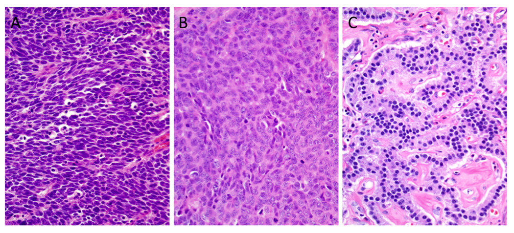
(A) Small cell neuroendocrine carcinoma (small cell NEC). The neoplasm is composed of small cells with minimal cytoplasm, fusiform nuclei, inapparent nucleoli, and a high mitotic rate. Apoptotic bodies are also frequent. (B) Compared to the carcinoma shown in A, this neoplasm contains large cells with abundant cytoplasm. The nuclei have an open chromatin pattern with prominent nucleoli. Abundant mitoses are also seen. (C) Well-differentiated neuroendocrine tumor. Note the small uniform nuclei with delicate chromatin. The neoplastic cells show a trabecular growth pattern in a background of hyalinized and vascular stroma (hematoxylin and eosin stains, original magnifications x200).