
Figure 2. Immunohistochemical labeling patterns for DAXX and ATRX.
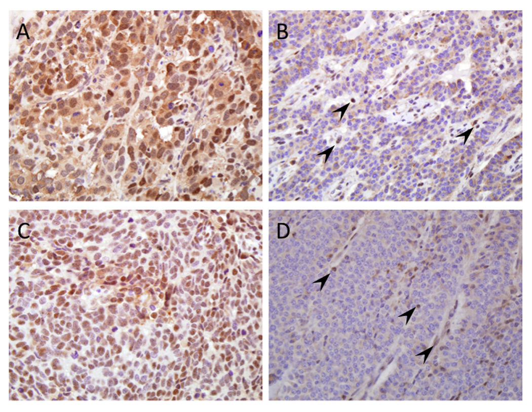
(A) Example of positive nuclear labeling for DAXX in a large cell neuroendocrine carcinoma. (B) Loss of nuclear labeling of DAXX in a well-differentiated neuroendocrine tumor. Endothelial cells and lymphocytes within the stroma are positive. (C) Positive nuclear labeling for ATRX in a small cell neuroendocrine carcinoma. (D) Loss of nuclear labeling of ATRX in a well-differentiated neuroendocrine tumor. Similar to that for DAXX shown in panel B, endothelial cells and lymphocytes within the stroma are positive (original magnifications x200).