
Figure 3. Immunohistochemical labeling for p53, Rb1 and Bcl-2 in small cell neuroendocrine carcinomas.
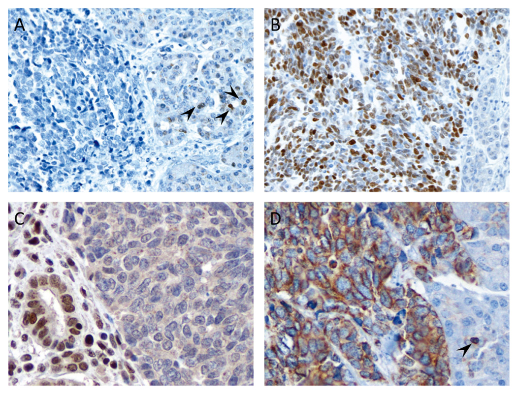
(A) Example of loss of nuclear labeling for p53. Scattered reactive acinar cells with positive nuclear labeling are present in the adjacent normal tissue (arrowheads). Sequencing analysis for TP53 in this patient revealed a nonsense mutation (p.E307X). (B) Example of diffusely positive nuclear labeling for p53. (C) Example of loss of nuclear labeling for retinoblastoma (Rb1) protein. Adjacent nonneoplastic cells show positive nuclear labeling (left side). (D) Diffuse cytoplasmic positivity for Bcl-2 protein. Note the reactive lymphocyte regarded as an internal positive control for this protein (arrowhead) (original magnifications x200).