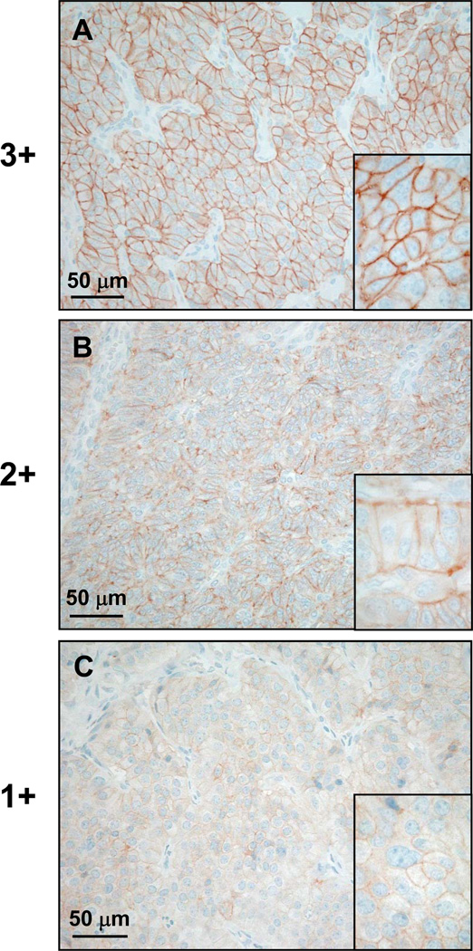
Figure 1.
Scoring of immunohistochemical staining intensity in typical examples (A: ileal neuroendocrine carcinoma, B: neuroendocrine carcinoma of the gall bladder, C: pulmonary carcinoid tumor). A: 3+ staining intensity represents strong staining at low magnification and fully circumferential staining of tumor cells membranes at high magnification (inset). B: 2+ staining intensity equals strong staining at low magnification, but no staining of the entire tumor cell circumference at high magnification (inset). C: 1+ staining intensity is defined by weak staining of the tumor cell membranes at low and high (inset) magnification.

