Figure 4.
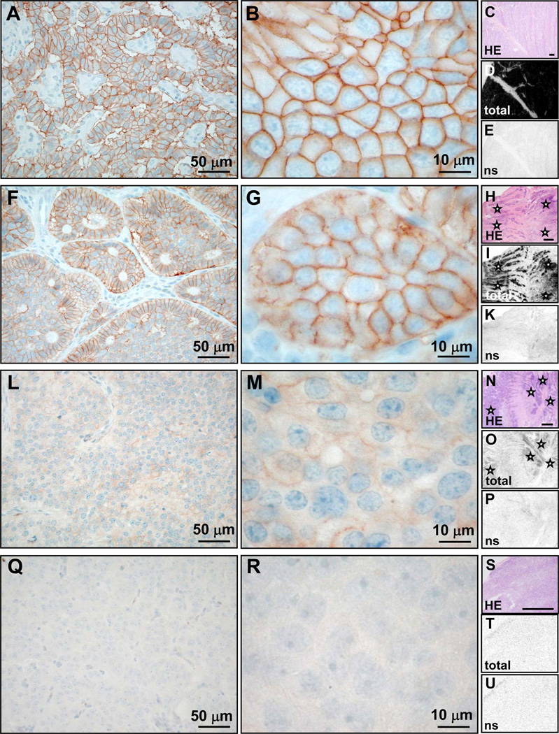
Comparison of UMB-1 immunohistochemistry (concentration 1:20, pre-treatment PC-C; first two columns) with 125I-[Tyr3]-octreotide receptor autoradiography (last column; bars indicate 1mm) in typical examples of tumors with high somatostatin receptor levels (rows 1 and 2), low somatostatin receptor levels (row 3) and no somatostatin receptor expression (row 4). A–E, malignant insulinoma: Strong membranous UMB-1 staining of most tumor cells (A) and often of entire tumor cell circumference (3+; B). Receptor autoradiography on serial tissue sections of the same case showing the tumor tissue stained with HE in C, very strong total 125I-[Tyr3]-octreotide binding to the entire tumor sample in D and complete displacement of 125I-[Tyr3]-octreotide by cold octreotide, providing evidence of specificity of somatostatin receptor binding in E (ns = non-specific 125I-[Tyr3]-octreotide binding in presence of excess cold octreotide). There is an excellent correlation between strong UMB-1 staining and high 125I-[Tyr3]-octreotide binding levels. F–K, ileal neuroendocrine carcinoma: Strong membranous UMB-1 staining of a large proportion of tumor cells (F), in some tumors cells affecting the entire tumor cell circumference (3+; G). This corresponds well to the strong specific 125I-[Tyr3]-octreotide binding to the entire tumor (asterisks) in the autoradiography experiment (H–K). L–P, ileal neuroendocrine carcinoma: Weak membranous UMB-1 staining at low (L) and high (M) magnification, correlating well with weak 125I-[Tyr3]-octreotide binding to the tumor tissue (asterisks; N–P). Q–U, benign insulinoma: No UMB-1 staining (Q, R), which matches well with negative receptor autoradiography (S–U).

