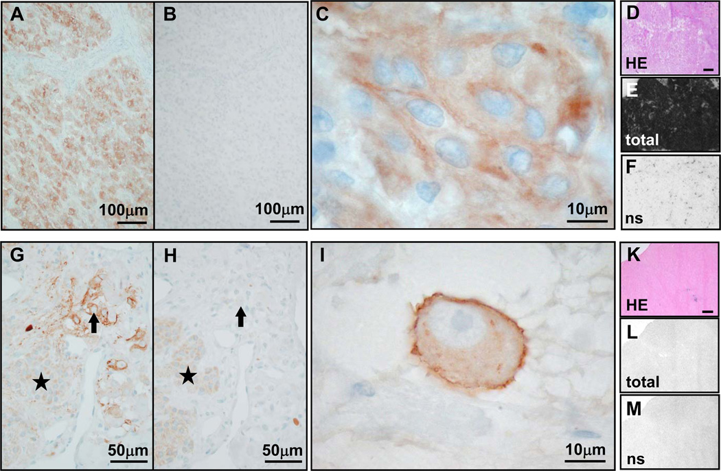
Figure 5.
Cases requiring cautious interpretation of immunohistochemical UMB-1 staining (concentration 1:20; pre-treatment PC-C; columns 1–3) in view of 125I-[Tyr3]-octreotide autoradiography results (last column; bars indicate 1mm). A–F, meningioma: Wide-spread and strong UMB-1 positivity at low magnification (A) that is completely abolished by the immunogen peptide (B). At high magnification (C), there is often a blurred staining in the area of the cell membrane, but only rarely a clear membranous staining. This staining pattern is probably due to prominent intercellular interdigitations. Autoradiographically, there is strong specific 125I-[Tyr3]-octreotide binding to the entire tumor tissue (D–F). Although <10% of tumor cells show membranous staining, this tumor is an excellent candidate for in vivo somatostatin receptor targeting. G–M, ganglioneuroblastoma: UMB-1 staining is present in single ganglionic tumor cells (arrow; G) in a membranous distribution (I, high magnification), but not in larger areas of neuroblastic tumor cells (asterisk; G). UMB-1 staining of ganglionic tumor cells is completely abolished by the immunogen peptide (H), providing proof of specific UMB-1 staining. No specific 125I-[Tyr3]-octreotide binding with receptor autoradiography (K–M). Despite strong tumor cell staining, this tumor is apparently not suitable for in vivo somatostatin receptor targeting, as the total receptor number per tumor mass is too low.

