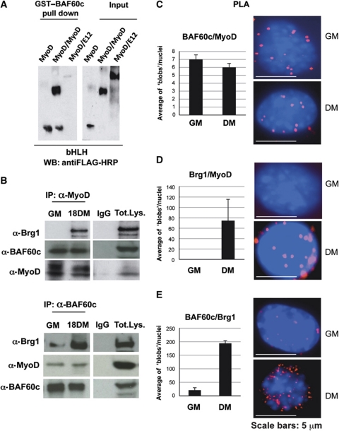
Figure 1.
BAF60c and MyoD interact in vitro and in vivo. (A) Pull-down assay was performed using GST–BAF60c and baculovirus-purified Flag-tagged MyoD, MyoD∼MyoD and MyoD∼E12 forced dimmers. (B) Co-immunoprecipitations from nuclear extracts of myoblasts and myotubes, with anti-MyoD and anti-BAF60c antibodies. (C–E) PLA was used to monitor nuclear ‘in situ’ interactions between MyoD, Brg1 and Flag–BAF60c during C2C12 differentiation. Each fluorescent dot, ‘blob’, represents the co-localization of the indicated proteins in myoblasts and myotubes. The quantification of the blobs is represented in the adjacent graphic. The BlobFinder software (Allalou and Wählby, 2009) was used to localize and quantify the blobs from images acquired with fluorescent microscopy. The average of blobs/nuclei in the graphic corresponds to the quantification of several images from three different experiments in myoblasts and myotubes.

