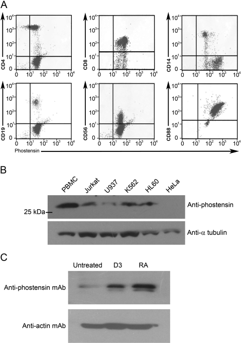
Figure 2.
Phostensin is expressed in leukocytes and in leukemic cell lines. (A) Leukocytes were double stained for cell surface markers (CD4, CD8, CD14, CD19, CD56, or CD88) and phostensin expression and analyzed by flow cytometry. (B) Phostensin is present in leukemic cell lines. Cellular proteins were extracted by 1% SDS and ultrasonication. An aliquot (100 µg) of each crude extract was analyzed by SDS-PAGE, transferred onto polyvinylidene difluoride membranes, and blotted with PT2 (1:1000). The arrow indicates phostensin recognized by PT2. (C) Differentiation of HL60 cells was induced by retinoic acid and 1,25-dihydroxyvitamin D3, respectively. Cellular proteins were extracted by 1% SDS and ultrasonication. All components of extracted cells were analyzed by Western blot using the PT2 monoclonal antibody.

