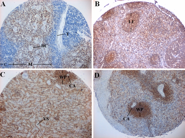
Figure 4.
Phostensin is abundant in the thymus, lymph nodes, and spleen. Tissue sections were examined by immunohistochemistry analysis with the PT2 monoclonal antibody. (A) Thymus. C, cortex; HC, Hassall’s corpuscles; M, medulla; T, trabeculae. (B) Lymph node. LF, lymphoid follicle. (C) Spleen. CA, central artery; VS, venous sinusoid; WP, white pulp. (D) Spleen. WP, white pulp; CA, central artery.

