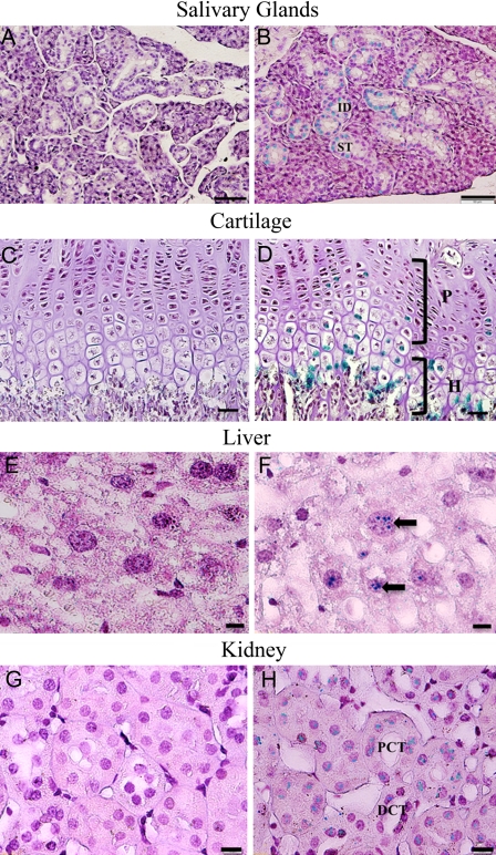
Figure 3.
Dentin sialophosphoprotein (DSPP)–lacZ expression pattern in 3-month-old homozygous Dspp knockout mice. The following expression patterns were observed: Salivary glands: (A) salivary gland samples from C57BL/6J wild-type mice were used as negative control; (B) the interlobular duct (ID) as well as the striated duct (SD) of the submandibular salivary gland of the Dspp knockout mice showed positive lacZ staining; bar = 50 µm. Cartilage: (C) growth plate cartilage control from C57BL/6J wild-type mice; (D) growth plate cartilage of Dspp knockout mice expressing positive stain for lacZ in the proliferative (P) and hypertrophic zones (H) of the cartilage; bar = 25 µm. Liver: (E) liver tissues from C57BL/6J mice were used as control and showed no reaction to lacZ staining; (F) in Dspp knockout mouse liver, hepatocytes (black arrow) exhibited lacZ expression; bar = 10 µm. Kidney: (G) samples from C57BL/6J mice were used as negative control; (H) Dspp knockout mice exhibited positive signals in the distal (DCT) and proximal convoluted tubule (PCT) of the kidney; bar = 10 µm.

