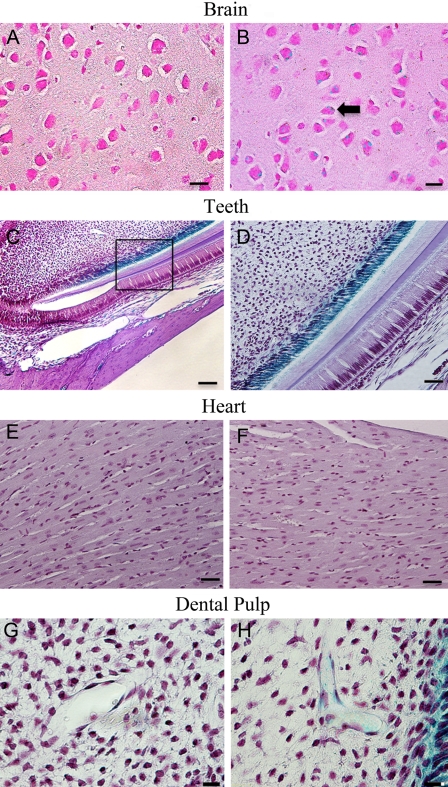
Figure 4.
Dentin sialophosphoprotein (DSPP)–lacZ expression pattern in 3-month-old homozygous Dspp knockout mice. The following expression patterns were observed: Brain: (A) cerebral cortex samples from the wild-type C57BL/6J mice used as control; (B), Dspp knockout mice showed bodies of neurons of ganglionic cell layer (black arrow) positively stained for DSPP-lacZ expression; bar = 25 µm. Teeth: (C) Dspp knockout mouse incisor exhibiting positive staining for DSPP-lacZ expression in the odontoblast layer of incisors; (D) higher magnification of the boxed area in C. The blue staining in the cytoplasm of odontoblast layer was possibly caused by the diffusion of β-galactosidase staining from the nuclei into the cytoplasm. Bar: C = 100 µm; D = 50 µm. Heart: no difference was observed between the wild-type C57BL/6J (E) and Dspp knockout (F) mice; bar = 25 µm. Dental pulp: (G) control sample from the wild-type mice; (H) LacZ signaling was noticed in the blood vessels in the dental pulp, particularly in the pericytes surrounding the blood vessels; bar = 10 µm.

