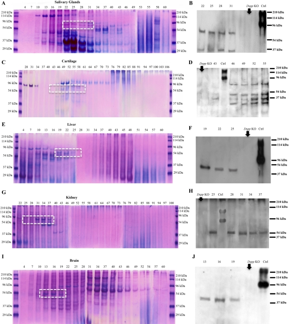
Figure 5.
Stains-all staining and Western immunoblotting in salivary glands (A, B), articular cartilage (C, D), liver (E, F), kidney (G, H), and brain (I, J). Numbers at the top of each image represent fraction numbers of the Q-Sepharose chromatography. (A, C, E, G, I) Stains-all staining for every third fraction in the chromatographic region of interest was performed to illustrate the existence of acidic proteins. The identity of dentin sialoprotein (DSP) in the corresponding fractions (dotted white boxed region) was confirmed by Western immunoblotting (B, D, F, H, J) using the anti-DSP-2G7.3 antibody. DSP was detected in fractions numbered on the top of the figures. The differences in the fractions in which DSP eluted may be attributed to the posttranslational modifications of DSP in these different tissues. The tissue extracts from Dspp knockout (Dspp KO) mice were used as negative controls (B, D, F, H, J). Ctrl: 0.3 µg of DSP isolated from incisor dentin of rat. Note that DSP extracted from these tissues migrated faster than that isolated from rat dentin (i.e., the positive Ctrl).

