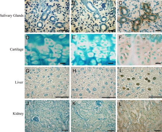
Figure 7.
Immunohistochemistry using the anti-DSP-2C12.3 monoclonal antibody (C, F, I, L). Mouse IgG of the same concentration as the anti-DSP-2C12.3 was used as the negative control (B, E, H, K). The tissues from Dspp knockout mice were also used as negative controls (A, D, G, J). DSP immunoreactivity was identified in the interlobular duct as well as striated duct of the submandibular salivary gland (C). In the cartilage, immunostaining was identified in the proliferative and hypertrophic zones (F). Dentin sialoprotein (DSP) immunoreactivity was also detected in the liver sac containing hepatocytes (I). In the kidney, immunostaining for DSP was widely distributed throughout the distal as well as proximal convoluted tubule (L). Bar: A-C = 50 µm; D-F = 10 µm; G-L = 50 µm.

