Figure 8.
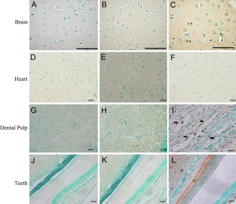
Immunohistochemistry using the anti-DSP-2C12.3 monoclonal antibody (C, F, I, L). Mouse IgG of the same concentration as the anti-DSP-2C12.3 was used as the negative control (B, E, H, K). The tissues from Dspp knockout mice were also used as negative controls (A, D, G, J). Dentin sialoprotein (DSP) immunoreactivity was identified in the cerebral cortex, and bodies of neurons of the ganglionic cell layer showed a positive signal for DSP expression (C). Heart showed no difference between the Dspp knockout mouse sample (D), mouse IgG control (E), and the anti-DSP antibody specimens (F). The blood vessels (BV) in the dental pulp showed positive immunostaining in the areas containing pericytes (I, arrows). Teeth sample, showing pulp (P) and dentin (D) region, from wild-type mice was used as the positive control (L). Bar: A-L = 50 µm.

