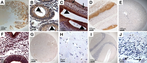
Figure 2.
Expression of ezrin in the alimentary system and the nervous system of human embryonic, fetal, and normal adult tissues. Ezrin-positive reactivity was defined as those showing brown spotting. (A) Ezrin was positive in adult intestine (40×). (B) Ezrin protein was mainly expressed in the brush border (triangles) of the absorptive cells of the gastrointestinal tract (200×). (C) Immunoreactivity for ezrin was present in epithelial cells (triangles) and smooth muscle cells (arrows) of the gastrointestinal tract at 8 weeks’ gestation (200×). Ezrin was expressed in adult esophagus (D, 40×) but was undetectable in liver tissues (E, 40×). (F) Medullary epithelium and all nerve cells showed significant expression of ezrin at 4 weeks’ gestation (200×). (G–H) Ezrin was undetectable in the nervous system of fetuses and adults. (G) Cerebrum (40×). (H) Nerve fibers and cells of the cerebrum (200×). (I) Cerebellum (40×). (J) Cells of Purkinje cell layer (arrows), granular layer, and molecular layer of the cerebellum (200×).

