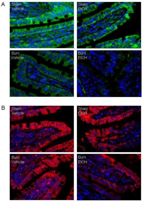
Fig. 1. Representative micrographs showing immunofluorescence localization of occludin and claudin-1 protein in small intestine one day after EtOH and burn injury.
Representative photomicrograph showing small intestine sections stained with anti-occludin antibody and Alexa Fluor® 488 conjugated goat anti-rabbit IgG (Panel A) or with anti-claudin-1 antibody and Texas Red® conjugated goat anti-rabbit IgG (Panel B). Nucleus was stained with Hoechst (blue). Localization of occludin (green) and claudin-1(red) were observed by fluorescence microcopy (x1000). Similar results were obtained in 3-4 animals in each group.

