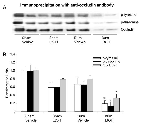
Fig. 2. Representative blots showing occludin protein expression and phosphorylation in small intestine mucosa one day after EtOH and burn injury.
Mucosal homogenates were immunoprecipitated with anti-occludin antibody and immunoblotted with phospho-antibodies (A). Band densities were quantitated by image analysis, normalized to the average of the band densities of sham animals and are shown as mean ± SEM from 3-4 animals in each group in Panel B.*, p<0.05 compared with other respective groups; #, p<0.05 compared with respective sham vehicle.

